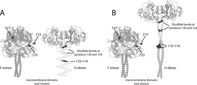
FIGURE 8.
Two possible hypotheses of envelope glycoprotein alignment. H stalk domains are represented in unknown (A) or helical (residues 58–122) (B) conformation. The helical H stalk places H residues 110–114 and F residues 233 and 219 at the lateral face of the prefusion CDV F trimer at an equal distance above the viral envelope, making short range interactions structurally conceivable (B). The ribbon models of the glycoprotein oligomers are aligned at their transmembrane domains. Helical modeling of H stalk residues 58–122 is based on the predictions of SSpro (54) (81% helical). Cysteines 139 and 154 engaging in intersubunit disulfide bonds (55) are shown.

