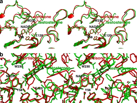
FIGURE 8.
Comparisons of steroid binding modes in catalysis. a, superposition of the AKR1D1·NADP+·cortisone complex (red) on the AKR1C9·NADP+·testosterone complex (green) showing that the A-ring of the substrate binds ∼1 Å deeper in the active site of the former enzyme relative to the position of the nicotinamide ring of NADP+. Presuming that NADPH binds similarly to NADP+, the 4-pro-(R)-hydride of NADPH would be adjacent to the substrate C-3 carbonyl in the AKR1C9 active site (green dotted lines), whereas the 4-pro-(R)-hydride of the NADPH cofactor would be adjacent to C-5 of the substrate carbon-carbon double bond in the AKR1D1 active site (red dotted lines). b, same orientation as a, but a close-up view showing all atoms.

