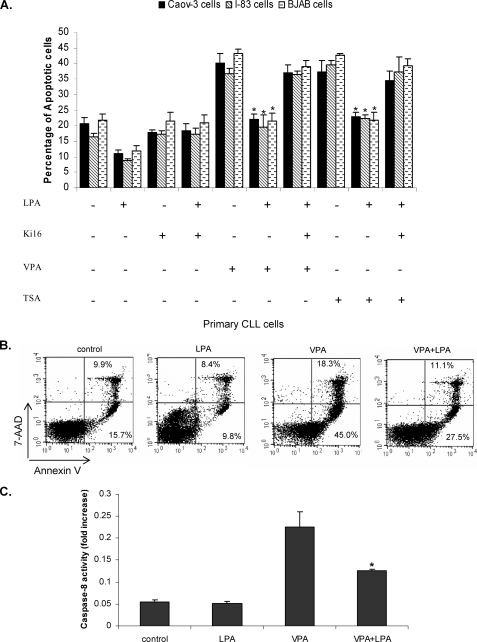
FIGURE 1.
LPA blocks HDAC inhibitor-induced apoptosis. A, I-83, BJAB, and CaOv-3 cells were pretreated with 1 μm Ki16425, 1 mm VPA, or 75 nm TSA, following 10 μm LPA treatment in serum-free medium. Apoptosis was determined by membrane permeability assay as described under “Experimental Procedures.” Data represent a trend in four independent experiments. B, primary CLL cells were treated with LPA and VPA, as in A, and apoptosis was measured by flow cytometry following annexin V and 7-AAD staining. Cells that were 7-AAD-negative and annexin V-positive were undergoing apoptosis. The percentage of cells in each quadrant is indicated in the quadrant. C, I-83 cells were treated with VPA alone or in combination with LPA. The 50-μg cell lysate was used for caspase-8 activation assay as described under “Experimental Procedures.” Data represent a trend in four independent experiments.

