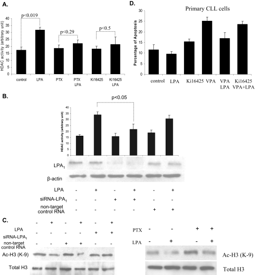
FIGURE 6.
LPA-induced HDAC activity was mediated via LPA1 receptor. A, I-83 cells were pretreated with 1 μm Ki16425 or 100 ng/ml PTX for at least 1 h before incubating with LPA. HDAC activity was measured in the nuclear extracts. B, HEK293 cells were transfected with siRNA-LPA1 for 48 h before treatment with LPA, and cell lysate was analyzed for HDAC activity. Values represent mean ± S.E. for three separate experiments. Transfection efficiency was assessed by immunoblotting experiments using rabbit polyclonal antibody anti-EDG2 (LPA1). β-Actin represents a loading control. C, protein level of acetyl histone H3 was examined by immunoblotting experiment from the same lysate as described above. I-83 cells were pretreated with 1 μm Ki16425 or 100 ng/ml PTX for at least 1 h before incubating with LPA. The nuclear lysate was analyzed by Western blotting to determine the level of acetyl histone H3 (Ac-H3). D, CLL cells were treated with 1 μm Ki16425, 1 mm VPA, or 75 nm TSA, or alone or in combination with 10 μm LPA in serum-free medium. Apoptosis was determined by membrane permeability assay as described under “Experimental Procedures.” Standard errors were determined from three independent experiments.

