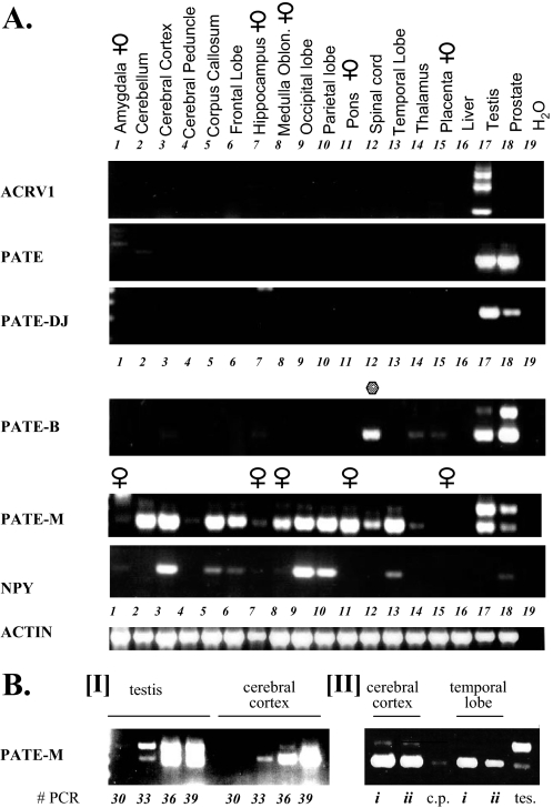
FIGURE 12.
Expression of human PATE-like genes in different regions of the brain. A, upper panel, cDNAs prepared from different individual regions of the brain as indicated, were subjected to RT-PCR analyses. Expression analysis of the human PATE-like genes was performed with forward and reverse primers that always spanned an intron and the observed RT-PCR product at all times corresponded to the size expected of a spliced mRNA. For the ACRV1, PATE, PATE-M, PATE-DJ, and PATE-B genes, the forward and reverse primers were located in the first and third exons (coding for the signal peptide and cysteines 6-10, respectively). In addition, cDNAs derived from testes and prostate on the one hand and liver and placenta on the other hand were also analyzed for expression of the PATE-like genes and served as positive and negative controls, respectively. PCR was performed for 35 cycles. Control for cDNA integrity was provided by the actin primers, as indicated, and expression analysis of the brain neuropeptide NPY served as a positive control for expression of a brain neuropeptide in specific regions of the brain. B, panel I, to assess the relative PATE-M expression levels in testis and cerebral cortex, a semiquantitative RT-PCR analysis was performed wherein samples were taken following 30, 33, 36, and 39 PCR cycles and subjected to agarose gel analysis. The fact that there are two differently sized PATE-M isoforms, each possessing a distinct duplex melting temperature, precluded real time PCR analysis for this gene. Panel II, to assess the generality of PATE-M gene expression in the brain, cDNA samples deriving from cerebral cortex and temporal lobe isolated from different individuals (i and ii) were subjected to PATE-M RT-PCR analysis. cDNAs prepared from testis (tes) and cerebral peduncle (c.p.) served as positive and negative controls, respectively.

