FIG. 9.
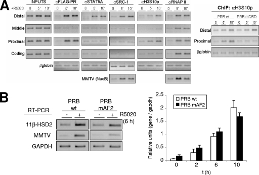
Coactivator recruitment and histone modifications at the 11β-HSD2 promoter. (A) TYML cells expressing FLAG-tagged WT PRB, cultured as described for Fig. 1A, were untreated (0) or treated with R5020 (10 nM) for 5 or 10 min, harvested, and used for ChIP experiments with anti-FLAG tag (αFLAG), STAT5A, SRC-1, H3S10p, and RNAP II antibodies. The precipitated DNA fragments were subjected to PCR analysis with specific primers corresponding to the indicated 11β-HSD2 promoter regions or MMTV nucleosome B and the β-globin gene as a control. For the right panel, ChIP with H3S10p antibody was performed on chromatin extracted from TYML cells expressing FLAG-tagged WT PRB or PRB-mDBD, untreated or treated with R5020. (B) TYML cells expressing WT PRB or PRB-mAF2, cultured as described for Fig. 1A, were untreated (0) or treated with R5020 (10 nM) for 2, 6, or 10 h. Cells were harvested, and total RNA was extracted. (Left) 11β-HSD2 and MMTV-Luc mRNA expression levels were analyzed by RT-PCR with specific primers. GAPDH cDNA-specific primers were used as a control. PCR products were run on a 1.2% agarose gel and visualized with ethidium bromide. (Right) 11β-HSD2 mRNA expression was analyzed by RT and real-time PCR with specific primers. GAPDH cDNA-specific primers were used as a control. The values represent the means and ranges of a representative experiment performed in duplicate, expressed as numbers of 11β-HSD2/GAPDH relative units.

