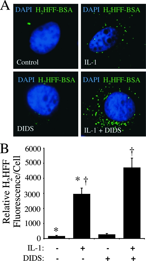
FIG. 2.
DIDS treatment of MCF-7 cells at the time of IL-1β stimulation increases intraendosomal ·O2−. Endosomal ·O2− production by MCF-7 cells was detected using H2HFF-BSA (50 μg/ml) loaded into the endosomal compartment following stimulation with IL-1β (50 ng/ml) for 20 min. (A) Representative fluorescent images of MCF-7 cells stained with H2HFF and DAPI following the treatments indicated in the lower-left corner of each panel. DIDS (500 μM) was added 10 min prior to IL-1β stimulation. The cells were fixed in 4% paraformaldehyde 20 min after IL-1β treatment and stained with DAPI (blue) for localization of the nucleus. The green fluorescence represents oxidized H2HFF resulting from ·O2− production. Images were taken with a 63× objective. (B) Quantification of H2HFF fluorescence for the conditions shown in panel A. The relative levels of fluorescence for the treatment groups (n = 5) were compared using one-way analysis of variance followed by the Bonferroni posttest (†, P < 0.05).

