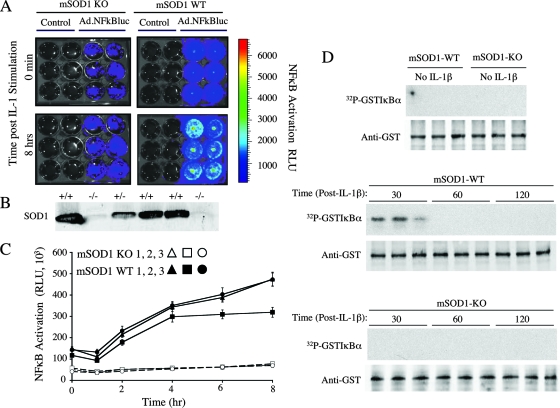
FIG. 8.
SOD1 is important for IL-1β-induced NF-κB activation. SOD1 knockout (SOD1-KO) and SOD1 wild-type (SOD1-WT) PMDFs were harvested as described in Materials and Methods and seeded in 12-well plates. Cells were infected with an NF-κB-inducible luciferase reporter carried on a recombinant adenoviral vector (Ad.NFκBLuc; multiplicity of infection, 300). Control wells were not infected. Luciferin (60 μg/ml) was added to the fibroblasts, and luciferase activity was assessed with a Xenogen IVIS imaging system. This was done prior to stimulation with IL-1β (50 ng/ml) and at several time points up to 8 h poststimulation. (A) Detection of NF-κB-inducible luciferase activity by the Xenogen IVIS imaging system. Representative wells are shown for SOD1 knockout and SOD1 wild-type PMDFs at 0 min and 8 h poststimulation. (B) Western blot for SOD1 protein in tissue from a litter of mouse pups used to derive PMDFs. Two of the six shown are knockouts (−/−). (C) Time course for NF-κB transcriptional activity following IL-1β stimulation. Values are luminescence (in relative light units [RLU] per well for each PMDF genotype (means and standard errors of the means; n = 6). (D) IKK assays for stimulation with IL-1β (50 ng/ml) at various time points. Samples were resolved by SDS-PAGE and transferred to nitrocellulose prior to autoradiography. Shown are results for three representative samples for each condition, including the control (unstimulated) and 30, 60, and 120 min after IL-1β stimulation. The top panel in each set shows a 32P-labeled GST-IκBα autoradiogram, and the bottom panel is a Western blot of the same filter with anti-GST antibody to control for sample loading.

