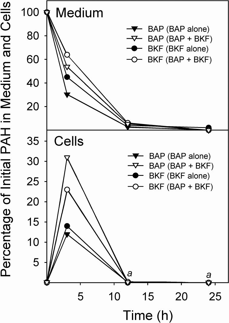
Fig. 4.
Uptake and metabolism of BAP and BKF, individually and in a binary mixture, in T-47D cells. Confluent cultures of T-47D cells in 6-well plates were exposed to medium (2 mL/well) containing BAP, BKF, or BAP plus BKF. At the indicated times, the media and cell monolayers were harvested, extracted, and analyzed for BAP and BKF by HPLC with fluorescence detection. The percentages of the initial BAP or BKF remaining in the medium (upper panel) and cells (lower panel) when 1.5 μM BAP was added alone (BAP alone), when 1.5 μM BKF was added alone (BKF alone), or when 1.5 μM BAP and 1.5 μM BKF were added together (BAP + BKF) at the time points, are shown. aIndicates that neither BKF nor BAP was detectable (<0.3% of the initial PAH amount) in any of the cell extracts at these time points.

