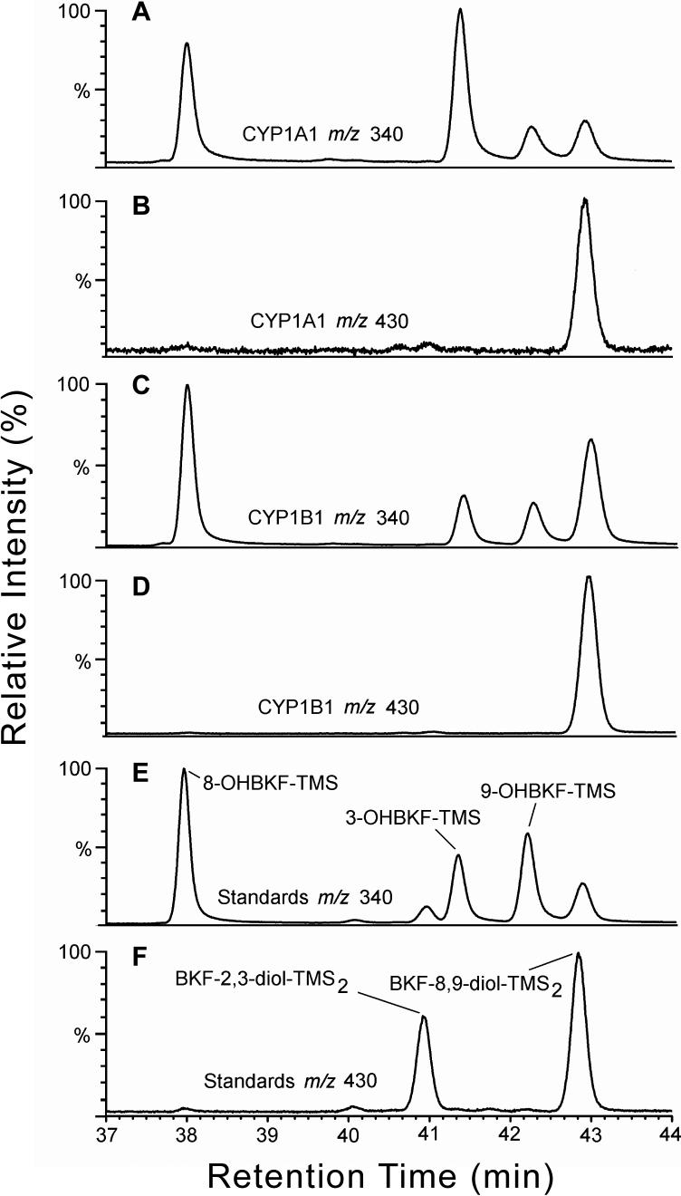
Fig. 7.
Analysis of microsomal BKF metabolism by GC/MS. After microsomal incubations (0.5-mL) with 10 pmol P450 and 10 μM BKF as substrate were extracted, TMS derivatives of the metabolites were prepared, and were analyzed by GC/MS together with BKF metabolite standards. Shown are selected-ion chromatograms for m/z 340 (A, C, and E), i.e., the value for the M+. ions of the TMS derivatives of OHBKFs, and for m/z 430 (B, D, and F) i.e., the value for the M+. ions of the TMS derivatives of BKF dihydrodiol metabolites, from the analysis of derivatized extracts of incubations with expressed CYP1A1 (A and B), CYP1B1 (C and D), and a derivatized standard mixture containing 3-, 8-, and 9-OHBKF-TMS, BKF-2,3-diol-TMS2 and BKF-8,9-diol-TMS2 (E and F).

