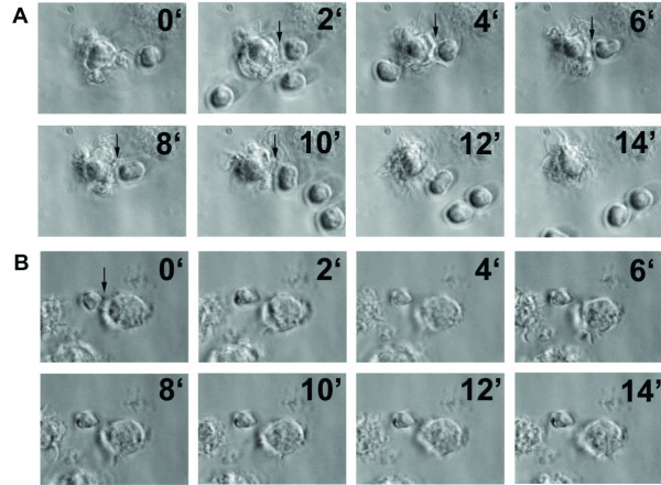
Figure 3.
Time-lapse microscopic analysis of the interaction between DCs and T cells after pre-incubation with P. yoelii-infected erythrocytes. DCs were differentiated in vitro and pre-incubated with uninfected (A) or P. yoelii-infected (B) erythrocytes before loading with OVA peptide 323–339. Naïve DO11.10 T cells that are specific for this OVA epitope were isolated from transgenic mice and added to DCs. Individual pictures frames from movies (Additional file 1 and Additional file 2) showing DC-T cell interactions. Arrows indicate contacts between DCs and T cells. Time in min is indicated in each frame. Representative results from one of five independent experiments are shown.

