Fig. 1.
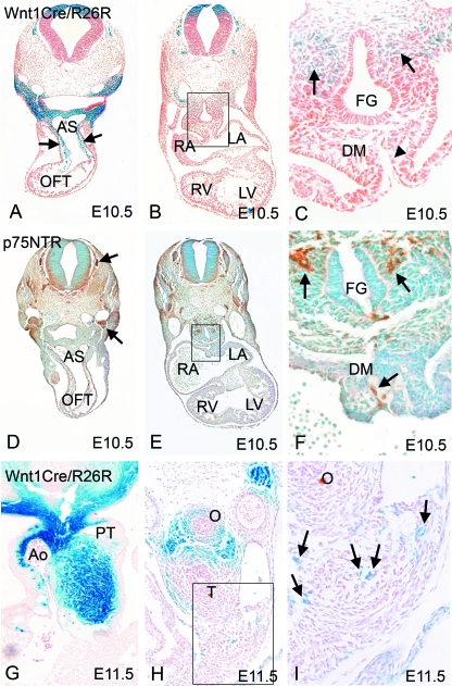
Neural crest cells arrive at the arterial pole before the venous pole. (A–C) Neural crest cells labelled blue by expression of β-gal using the Wnt1-cre/ROSA 26R system, are abundant in the outflow tract by E10.5 (arrows in A), but are absent from the dorsal mesocardium at this stage (arrowhead in C). Neural crest cells can be seen in the mesenchyme surrounding the foregut at this stage (arrows in C). (D–F) p75 NTR staining (brown) is absent from the branchial arches, aortic sac and outflow tract cushions at E10.5, but is found in the dorsal root and cranial ganglia (arrows in D). p75 NTR is also found in the dorsal mesocardium and in the mesenchyme surrounding the foregut (arrows in F). AS, aortic sac; DM, dorsal mesocardium; FG, foregut; LA, left atrium; LV, left ventricle; O, oesophagus; RA, right atrium; RV, right ventricle; T, trachea. (G) Neural crest cells are abundant in the outflow region at E11.5. (H,I) Only a few isolated neural crest cells (arrows in I) can be seen in the dorsal mesocardial connection at E11.5.

