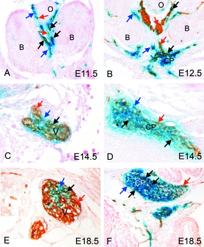
Fig. 2.
Neural crest cells contribute to the vagal nerve and the posterior cardiac plexus at the venous pole of the heart. In each case, blue arrows mark solely β-gal-stained cells (blue), red arrows mark solely NF160D-stained cells (brown) and black arrows marked double labelled cells (blue/brown). (A) By E11.5, double labelling for β-gal (blue; neural crest-derived cells) and NF160D (brown; neurons) shows that the vagal nerve is forming in the foregut mesenchyme and that this contains a mixture of brown (red arrows), blue (blue arrows) and double labelled (black arrows) cells. (B) At E12.5, the vagal nerve has differentiated further and can be seen to comprise mainly neural cells of non-neural crest origin (brown cells; red arrows). There is a smaller contribution of neural crest-derived neural cells (double labelled; black arrows) and neural crest cells of non-neural fate (blue cells; blue arrows). By this stage the cardiac plexus can also be seen. This also comprises cells of mixed origin (see above), although in this case the majority of cells derive from the neural crest (stained blue). (C,E) The vagus nerve continues to have neurons of neural crest and non-neural origin at E14.5 and at E18.5, with the majority being of non-neural crest origin (brown). (D,F) In contrast, the majority of neuronal cell bodies in the posterior cardiac plexus are of neural crest origin (blue). B, bronchus; CP, cardiac plexus; O, oesophagus; V, vagal nerve.

