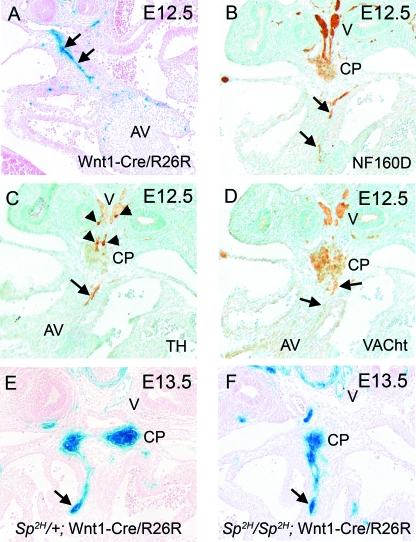
Fig. 3.
At E12.5, the majority of cells in the vagal nerve and posterior cardiac plexus are cholinergic parasympathetic neurons. (A) β-Gal staining reveals a stream of neural crest-derived cells (blue) within the dorsal mesocardial connection, which is reminiscent of nerve tracts (arrows). (B) NF160D staining marks the vagal nerve, the posterior cardiac plexus and confirms the presence of neuronal staining within the dorsal mesocardium (arrows). (C) The sympathetic neuron marker tyrosine hydroxylase marks only a subset of the cells in the vagal nerve and cardiac plexus (arrowheads), but does stain the nerve within the dorsal mesocardium. (D) In contrast, VAChT appears to stain the majority of the vagal nerve and posterior cardiac plexus and also localizes to the nerve in the dorsal mesocardium (arrows). (E) In Sp2H/+ embryos, which display a phenotype comparable with wild-type embryos, the stream of neural crest-derived cells resembling nerve tracts can be seen entering the heart through the dorsal mesocardium at E13.5 (arrow). Neural crest-derived cells are also observed in the vagal branches and cardiac plexus. (F) In Sp2H/Sp2H embryos at E13.5, similar patterns of β-gal staining are observed, indicating that the vagus nerve and cardiac plexus are formed normally and that cardiac innervation through the dorsal mesocardium occurs (arrow). AV, atrioventricular cushions; CP, cardiac plexus; V, vagal nerve.

