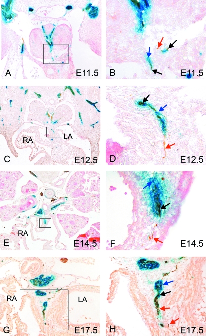
Fig. 4.
Nerves growing into the venous pole of the heart are a mixture of neural crest- and non-neural crest-derived neurons. In each case, blue arrows mark solely β-gal-stained cells (blue), red arrows mark solely NF160D-stained cells (brown) and black arrows marked double labelled cells (blue/brown). (A,B) Neural crest cells expressing β-gal (blue arrows) can first be seen in the dorsal mesocardium at E11.5, co-localizing with neuronal staining (red arrows). (C–H) Nerves can be identified within the venous pole of the heart throughout gestation, in each case comprising a mixed population of neural crest-derived neurons (black arrows), neural crest-derived non-neural cells (blue arrows) and more proximally, non-neural-crest derived neurons (red arrows). LA, left atrium; RA, right atrium.

