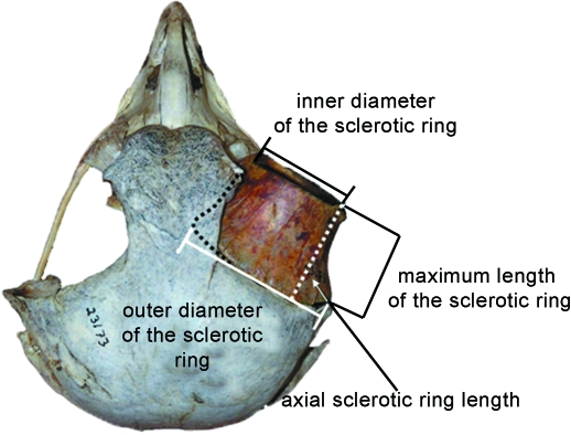
Fig. 1.
A superior view of a Nyctea scandiaca (snowy owl) skull. On the right the sclerotic ring is present in the orbit; on the left it is absent. The sclerotic ring houses that portion of the eye that protrudes from the orbit. The cornea protrudes laterodistally from the sclerotic ring, and that portion of the sclera that is contained within the orbit proper protrudes proximally. The four measurements taken on the sclerotic ring are here depicted: the inner diameter of the sclerotic ring (the bony correlate of corneal diameter), the maximum length of the sclerotic ring and the axial sclerotic ring length (both bony correlates of that portion of eye that protrudes from the orbit), and the outer diameter of the sclerotic ring. The axial sclerotic ring length was calculated using Pythagoras’ theory, with the maximum length of the sclerotic ring as the hypoteneuse of a right-angled triangle and half the inner diameter of the sclerotic ring subtracted from the inner diameter of the sclerotic ring as the base. Solving for the remaining side yields the axial sclerotic ring length.

