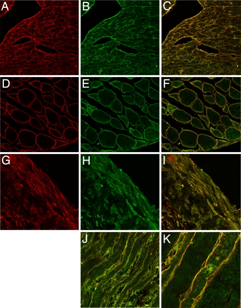
Fig. 2.
Colocalization of Bves and GEFT in cross and transverse sections of mouse cardiac, skeletal, and smooth muscle. Bves, shown in red (A, D, and G), is primarily distributed at the cell periphery in cardiac (A–C, J), skeletal (D–F, K), and smooth muscle (G–I). GEFT (B, E, and H) also has distribution at the cell membrane in these muscle types, but it displays a broader intracellular localization at the myofibrils. Merged images are shown in C, F, I, J, and K.

