
Figure 4. Regulation of Bcl-2-family protein expression by IL-6, IFN-α or IGF-1 on XG-6 myeloma cells.
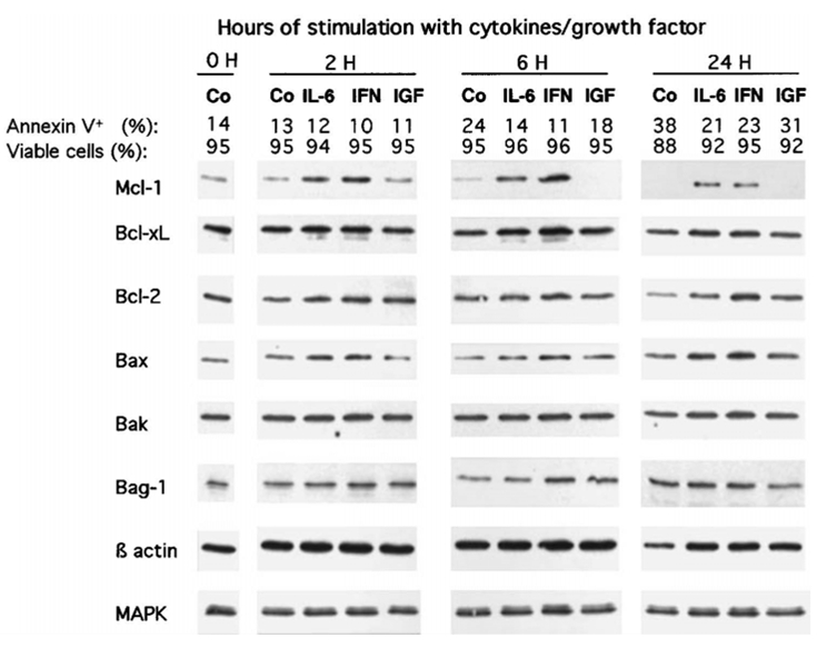
XG-6 myeloma cells were IL-6 and FCS starved for 1 hour and cultured for 2, 6 and 24 hours with no cytokine (Co), 2 ng/mL IL-6 (IL-6), 200 U/mL IFN-α (IFN-α) or 100 ng/mL IGF-1 (IGF) in RPMI1640 and 1% BSA. At the end of the culture, the cell count and the percentage of viable cells were assayed by trypan blue exclusion. The percentages of apoptotic cells were determined by staining with FITC-annexin V. Cells were immediately lysated and assayed for Bcl-2-family protein expression using Western blot analysis. The amount of proteins was carefully determined and 25 μg of total proteins from each culture group were loaded on the gel. In the experiments, MAPK expression was used as a loading protein control because it was not affected by the level of apoptosis. Western blots are for one representative experiment out of 3.