
Figure 1.
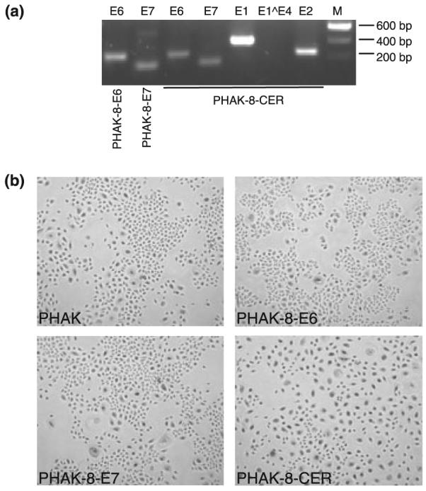
HPV transcripts expressed in human adult primary keratinocytes and morphology of cells transdused with different HPV8 genes. (a) Detection of HPV8 early gene transcripts in PHAKs infected with recombinant retroviruses. cDNA was amplified with primer for E6 and E7 to verify the presence of transcripts in PHAK-8-E6 (lane 1) and PHAK-8-E7 (lane 2) cells. These and also the primers for E1, E2 and E1∧E4 were used to show the presence of all early genes of HPV8 in PHAK-8-CER except for E1∧E4 (lanes 3–7). (b) Morphology of PHAK cells infected with recombinant retroviruses. Phase contrast photographs of PHAK, PHAK-8E6, PHAK-E7 and PHAK-8-CER at passage 2 are shown. Magnification, ×100.