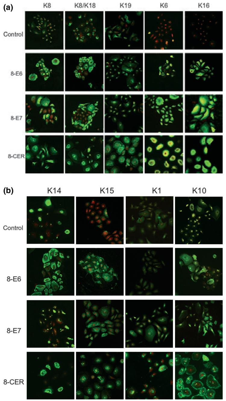
Figure 2.

Keratin immunofluorescence of PHAK, PHAK-8-E6, PHAK-8-E7 and PHAK-8-CER cell lines. Keratin staining, which fluoresces green, was detected using the TSA Fluorescein System. Propidium iodide (PI) staining, which fluoresces red, highlights DNA stain in the nuclei. The apparent yellow colour present in the K6 of the HPV8-CER cells was the result of a very intense fluorescence that sometimes obscures the red staining of the nuclei.
