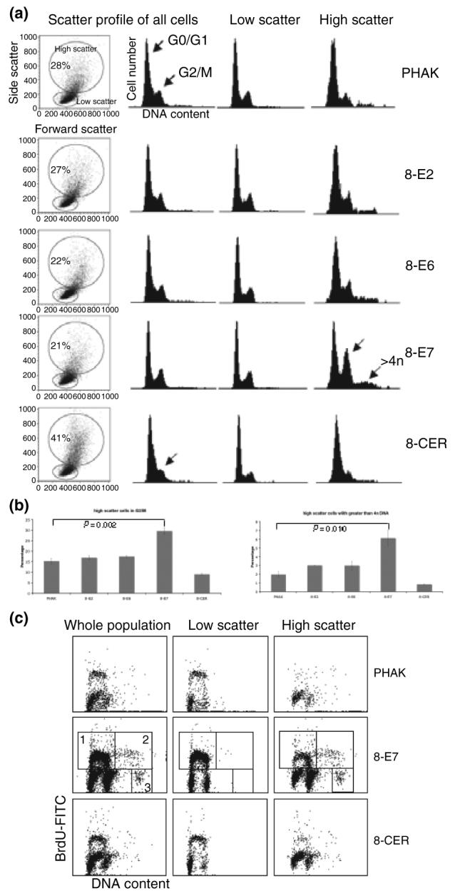
Figure 3.

Flow cytometric analysis of PHAKs expressing HPV8 early genes. (a, b) Relative DNA content of cells was determined by staining living cells with Hoechst 33342. Results shown are representative of at least three independent experiments. (c) BrdU incorporation into PHAK expressing HPV8 early genes. Box 1: 2n to 4n cycling cells; box 2: 4n to 8n cycling cells; box 3: octaploid cell population.
