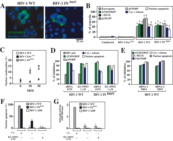
Figure 5. Activation and contribution of ATM to syncytial apoptosis of HIV-1-infected CD4+cell lines.
A–D. CD4+ HeLa cells were infected with HIV-1 WT, HIV-1 EnvVSV and HIV1 IND64V at a MOI of 10 for 38 h and then stained for ATMS1981P (A) or all other markers indicating syncytial apoptosis. The percentages (X±SD, n = 3) of syncytia exhibiting the indicated characteristics were quantified at an MOI of 10 (B), and the percentage of nuclei with apoptotic chromatin condensation (usual within syncytia) was quantified at different MOI (C). CD4+ HeLa cells were either transfected with specific siRNAs depleting ATM or treated with the ATM inhibitor KU-55933. Then, cells were infected with HIV-1 WT or HIV1 IND64V at a MOI of 10 for 48 h, and the expression of the viral protein p24, the activation of ATM or p53, and apoptosis were quantified (D). Results are plotted as the percent inhibition of the indicated parameters determined for syncytia (excluding single cells) compared to control siRNA transfected or untransfected cells. E. Effect of NBS1 knockdown on syncytial apoptosis of HIV-1 infected CD4+ HeLa cells. CD4+ HeLa cells were transfected with NBS1-specific or control siRNAs, and infected for 48h with HIV-1 WT (MOI of 10), followed by quantification of the indicated parameters by immunofluorescence. F, G. Effect of ATM inhibition on syncytial apoptosis in HIV-1 infected CEM T cells. Cells were infected with wild type HIV-1 in the presence (or in the absence) of the ATM inhibitor KU-55933, and nuclear apoptosis was determined 72 h later (F). Moreover, the number of infectious viruses contained in the culture supernatant was determined (G). Asterisks in F and G indicate significant inhibitory effects of KU-55933.

