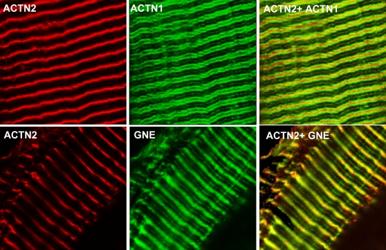
Figure 7. GNE and α-actinin 1 are expressed in stretched mouse muscle.
Confocal microscopy of stretched mouse spinalis muscle stained with antibodies recognizing the Z line marker α-actinin-2 (ACTN2, red), α-actinin-1 (ACTN1, green), and GNE (GNE, green). α-Actinin-1 antibodies(3A2) and GNE antibodies show a distinct but overlapping staining pattern: GNE Abs recognize a diffuse band centered on the Z line while α-actinin-1 Abs stain two distinct bands on either side of the Z line. Both Abs recognize also, as a fainter staining, the sarcomeric M line. Overlap stain between α-actinin-1 and α-actinin-2 Abs is minimal.

