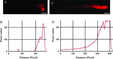
Fig. 2.
The FM4-64 staining in pollen tubes of Torenia fournieri. (A, C) FM4-64 fluorescence images of pollen tubes cultured without and with IAA treatment for 2 h, respectively, showing the FM4-64 staining signal focused at the tube apex and extended to the subapical region with gradually lower intensity. (B, D) Pixel values along the longitudinal axes of the tubes in (A) and (C), respectively, showing the significant increase of fluorescence intensity in IAA-treated tubes. Bars = 15 μm (A), 12 μm (C).

