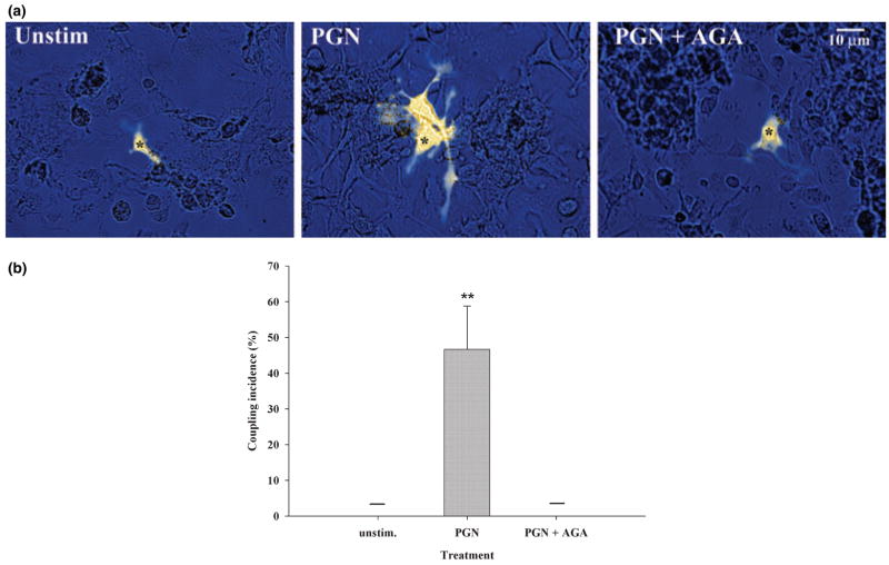
Fig. 4.
PGN treatment establishes functional gap junction communication in microglia. (a) Primary microglia were stimulated with 10 μg/mL PGN for 48 h, and gap junction communication was examined by single-cell microinjections of the gap junction-permeant dye LY CH. The gap junction-dependent spread of LY in activated microglia was confirmed by incubating PGN-treated cells with the gap junction inhibitor AGA (25 μM) before injections. Microinjected cells are denoted with asterisks. (b) Enumeration of the incidence of gap junction coupling revealed that approximately 50% of primary micro-glia stimulated with 10 μg/mL PGN for 48 h exhibited gap junction-dependent dye transfer to at least one neighboring cell. The horizontal bars representing results for unstimulated and PGN + AGA treatments indicate that microglia in these groups consistently failed to demonstrate any evidence of gap junction-dependent communication. Values are mean ± SD; the data presented are representative of six independent experiments evaluating at least 10 injected cells per treatment group per experiment. **p < 0.001 versus unstimulated microglia (Student’s t-test).

