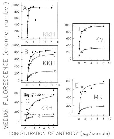
Figure 2.
Antibody affinity and competition assays. Binding of Pgp-specific mAbs was carried out for 30 min at 37°C and analyzed by indirect immunofluorescence. Each graph shows the median fluorescence for the cell population as a function of the amount of mAb added. (A) Binding of KK-H cells to mAb MRK16 in the absence (○) and in the presence (•) of 10 μM of vinblastine. (B) Binding of KK-H cells to mAb UIC2 in the absence of drugs (○), in the presence of vinblastine (•), and in the presence of 1 μM of oligomycin (▵). (C) Binding of KK-H cells to combinations of mAbs UIC2 and MRK16. Cells were preincubated 45 min at 37°C with the amounts of UIC2 indicated on the x axis, in the presence or in the absence of vinblastine. Cells then were washed and analyzed by indirect immunofluorescence either immediately (○ in the absence of vinblastine; • in the presence of vinblastine), or after incubation for 30 min at 37°C with 3 μg of MRK16 (□, no vinblastine; ▪, cells preincubated with vinblastine) or UIC2 (▵, no vinblastine). (D) Titration of KM-H with mAb UIC2 in the absence (○) and in the presence (•) of 10 μM of vinblastine. (E) Titration of MK-H with mAb UIC2 in the absence (○) and in the presence (•) of 10 μM of vinblastine.

