
Fig. 6. Schematic of astrocyte processes interacting with synapses and one blood vessel (red circle).
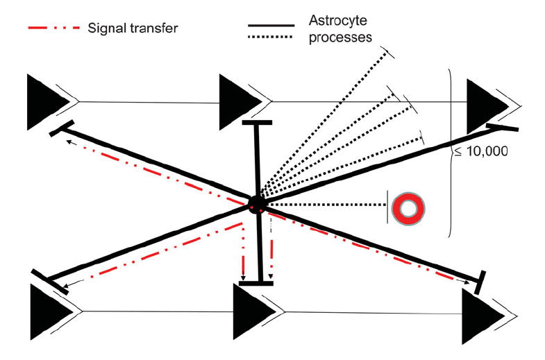
The black circle in the center represents an astrocyte cell body whose processes extend to many synapses (≤10 000). Shown are six synapses in two parallel neuronal circuits. Dashed black lines show members of part of the extensive astrocyte process domain in other planes. The postulated information pathway between astrocyte processes and some of the synapses of the same circuit, or between parallel synapses, is shown as a dashed red line. Astrocytes do not fire action potentials, so the signal is viewed as a spread of either intracellular Ca2+ or another intracellular messenger or messenger system. It is unlikely to be an electrotonic spread of a local depolarization based on calculations of the length constant of astrocytes in situ (Trachtenberg and Pollen, 1970). It is proposed that the perisynaptic astrocyte processes serve to maintain the local environment around the synapses for optimal functioning by localizing key proteins involved in uptake of, for example, glutamate and H+ and channels or transporters for K+ clearance released at the active synapse, and the activity of these fluxes is controlled by local feedback loops. These two possibilities (synaptic homeostasis based on feedback versus an instructional role based on initiating and integrative functions) are extremely difficult to distinguish either in situ or in vivo and, so far, there seem to be insufficient reasons to postulate the latter. See text for a fuller discussion.