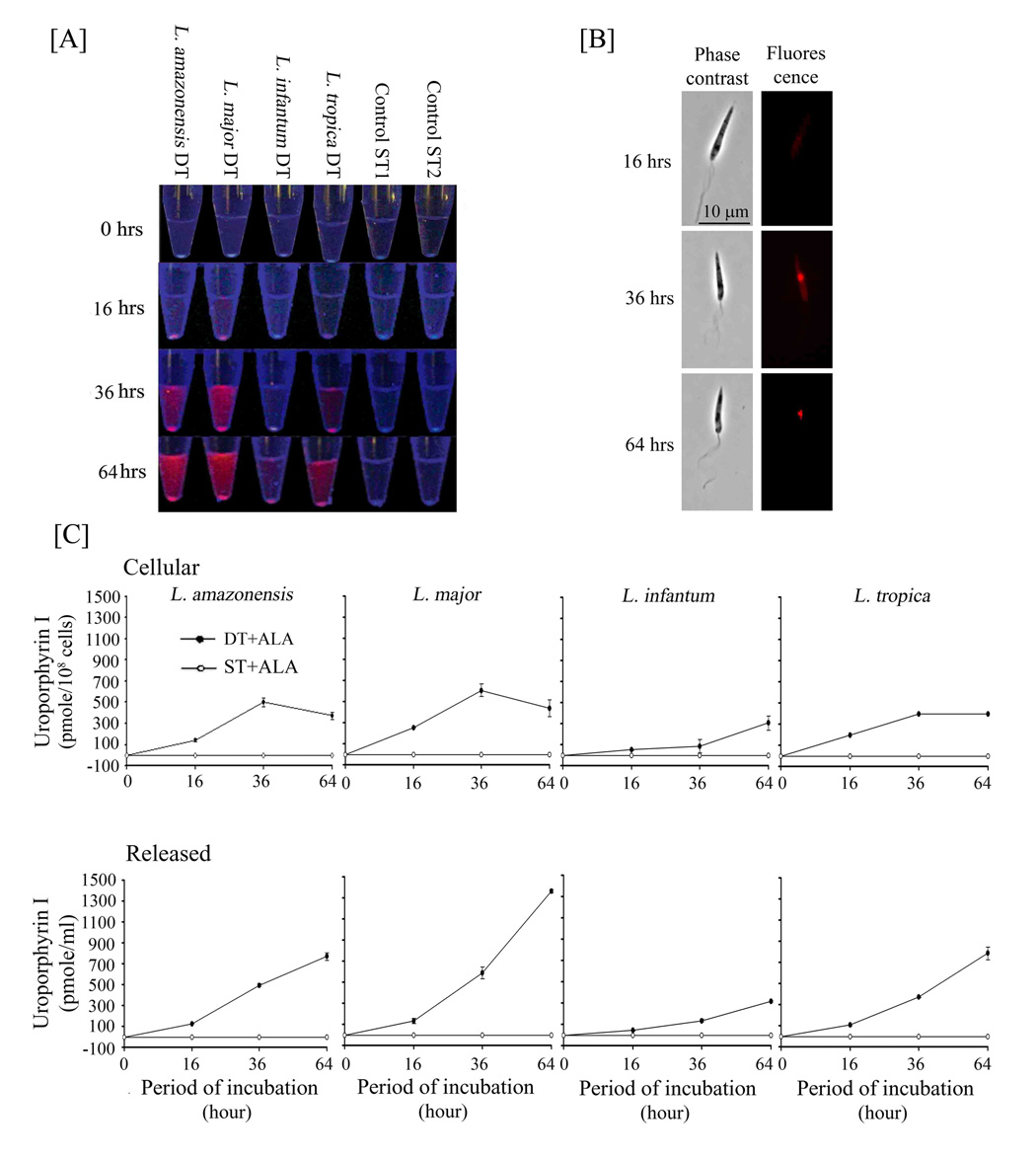
Fig. 4. Kinetics of delta-aminolevulinate-induced cellular neogenesis of uroporphyrin I, its cellular distribution and extracellular release.
[A] Double transfectants (DT) of all four Leishmania species grown to stationary phase were exposed to 1 mM ALA under non-growing conditions for up to 64 hr as described. Samples were withdrawn at the indicated intervals after ALA exposure and centrifuged to separate supernatants and cells. All samples were examined for fluorescence intensity under a UV lamp immediately [A] and for cellular distribution by fluorescent microscopy [B] [using excitation 405 nm (D405/10X), dichroic 485 nm (485DCXR) and emission 610 nm (RG610LP) filter set from Chroma USA] (L. major shown as a representative), and subsequently solvent-extracted for porphyrin quantitation by fluorimetry (400 nm excitation and 600 nm emission wavelengths) (DT+ALA versus ST+ALA) [C]. Concentrations of cellular and released uroporphyrin I were estimated against a standard curve of uroporphyrin I (see Legend to 3B). Note: Cellular emergence (starting from ~16 hrs) and accumulation of uroporphyrin via vacuolar condensation (after ~24 hrs) and its increasing release with time (16–64 hrs) after exposure to ALA (except for a notable delay in L. infantum). See legend to Fig. 3A for ST1 and ST2 single-transfectants included in [A] and [C] as aporphyric controls.

