
Figure 4.

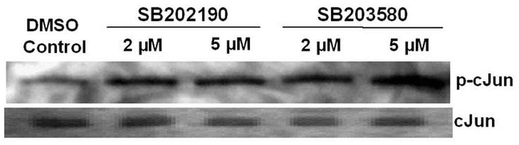
A. ATF-2 is phosphorylated by the p38 MAPK-specific inhibitor SB202190 A549 cells were treated with indicated concentrations of SB202190 or SP600125 for 16 hours. Anisomycin (1 µg/mL) was used as a positive control for ATF-2 phosphorylation. Cells were harvested after 16 hours of treatment; nuclear extract was prepared as mentioned in the Methods section. Nuclear extract (10 µg) was analyzed by Western analysis for phospho Thr71 using specific antibodies. Lower panel: Nuclear extracts were analyzed for unphosphorylated ATF-2 by Western analysis. Lane 1, untreated DMSO control cells; lanes 2–6, increasing concentrations of SB202190; lane 7, cells treated with only SP600125 (20 µM); lane 8, cells treated with 20 µM SP600125 and 1 µM SB202190; lane 9, cells treated with anisomycin (1 µM). (B) Phosphorylation of cJun by SB202190 or SB203580. A549 cells were treated with either 2 or 5 µM each of SB202190 or SB203580 for 1 hour. Cell lysates were prepared and JNK kinase assay was performed as described in the Methods section; lower panel, unphosphorylated cJun.