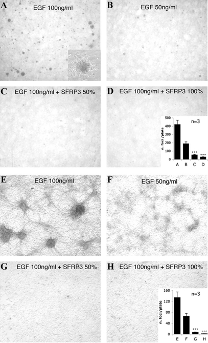
Figure 3. sFRP-3 inhibits proliferation EGF-dependent foci formation.
(A–D) sFRP-3 inhibits the EGF-induced colony formation and focal transformation of EGF-receptor over-expressing NIH 3T3 cells. EGF-dependent cell growth in soft agar. NIH3T3 clone 17 cells suspended in agar containing normal medium and treated with either 100 ng/ml (A) or 50 ng/ml (B) of EGF formed clearly visible colonies. The effect of EGF was inhibited in cells suspended in agar containing a 1∶1 mixture of normal medium and sFRP-3-containing medium (CM) (C) or the sFRP-3-CM alone (D). Photographs were taken after 20 days of growth at 2× magnification. Insert in D shows a 20× magnification of one colony; insert in H shows the results obtained by counting the colonies in 3 independent, reproducible experiments±SE. Error bars represent s.e.m. Triple asterisks, P<0.001 vs EGF. (E–H) EGF-dependent focal transformation. NIH3T3 clone 17 cells cultured for 10 days in normal medium containing 100 ng/ml EGF (E) or 50 ng/ml EGF (F) formed foci. This focal transformation was inhibited incubating the cells in a 1∶1 mixture of normal medium and sFRP-3-CM (G) or the sFRP-3-CM alone (H). Photographs were taken after 10 days of growth at 10× magnification. Insert in H shows the results obtained by counting foci in 3 independent, reproducible experiments±SE. Error bars represent s.e.m. Triple asterisks, P<0.001 vs EGF.

