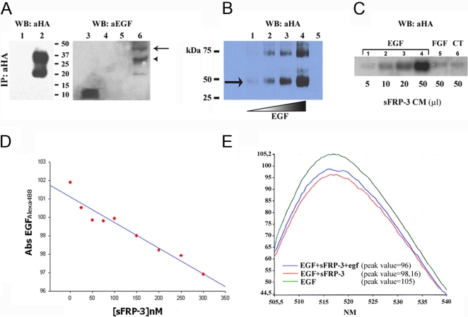
Figure 9. sFRP-3 binds EGF in vitro.
(A) Co-IP of sFRP-3-HA and EGF. Supernatant from 293 cells transfected with control plasmid (lanes 1, 4) or purified sFRP-3-HA (lanes 2, 5, 6) were incubated with mouse recEGF (lanes 4, 6), DTSSP cross-linked and analysed under non-reducing conditions. Western blot analysis with anti-HA (lanes 1, 2) and anti-EGF antibodies (lanes 3–6) revealed the presence of EGF bound to sFRP-3-HA (lane 6), but not in the control sample (lane 4). Note that the shift in molecular weight of the band(s) corresponding to sFRP-HA (lane 2) is due to EGF binding (arrow and arrowhead in lane 6). Free mouse recEGF was used as positive control for the immunoblot (lane 3). (B) Dose-dependent binding of EGF to sFRP-3. In vitro binding of increasing amount of EGF (100 ng, 200 ng, 400 ng and 600 ng,) to sFRP-3-HA (100 ng), followed by DTSSP crosslinking and revealed by Western blot analysis with anti-HA. The amount of EGF/sFRP-3 complexes (lanes 1–4) correlates with the increasing amount of EGF. As control for binding specificity, 100 ng of sFRP-3-HA alone (lane 5, not detectable) were cross-linked and revealed as before. (C) In vitro binding of sFRP-3 to EGF-loaded heparin beads. Western blot analysis with anti-HA on proteins adsorbed on EGF-loaded (lanes 1–4), FGF8-loaded (lane 5) and control beads (lane 6), following incubation with increasing amount of sFRP-3-HA supernatant (5–50 µl). sFRP-3 binds specifically to EGF, in a dose dependent manner. (D) Binding affinity of EGF for sFRP-3. EGFAlexa488 fluorescent absorbance at emission peak (517 nm) decreases in relation of increasing amount of sFRP-3. (E) Competition assay. Absorbance scanning from emission value 505 nm to 540 nm of EGFAlexa488 (green curve). A decreasing of the emission peak (517 nm) is shown in relation to EGFAlexa488/sFRP-3 interaction (red curve). By adding native egf to EGFAlexa488 prior to the incubation with sFRP-3, the absorbance reduction of the emission peak was smaller (blue curve).

