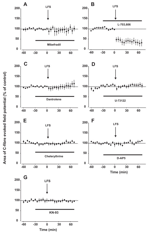
Figure 2.
Signalling pathways underlying the induction of LFS-induced LTP in vivo. In all graphs, area of C-fibre evoked field potentials (% of control) is plotted against time (min). Data are expressed as mean ± SEM (n = 5 for all groups). Spinal superfusion at the recording segment with the T-type VDCC blocker mibefradil (5 mM, A), the ryanodine receptor blocker dantrolene (500 μM, C), the PLC inhibitor U-73122 (500 μM, D), the PKC blocker chelerythrine (800 μM, E), the NMDA receptor blocker D-AP5 (100 μM, F) or the CaMKII blocker KN-93 (400 μM, G) fully blocked LTP induction by LFS. Blockade of spinal NK1 receptors with L-703,606 led to induction of LTD rather than LTP by LFS (B). See also Table 1.

