Figure 3.

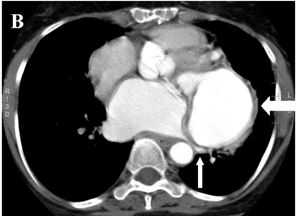
Axial source image of CT angiography showing the aneurysm (thick white arrow) compressing the left lower pulmonary vein (thin white arrow).

Axial source image of CT angiography showing the aneurysm (thick white arrow) compressing the left lower pulmonary vein (thin white arrow).