
Figure 4. Co-expression of two plasmids.
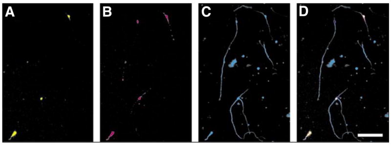
Green fluorescent protein (GFP) and the cell adhesion molecule L1 were cotransfected into cerebellar granule neurons (CGNs). The neurons were kept in culture for 48 h before being fixed and stained. (A) GFP fluorescence, (B) L1 immunofluorescence, and (C) β-tubulin immunofluorescence were used to determine transfection efficiencies. (D) A merge of the three channels in panels A, B, and C. Note that there are three cells expressing GFP and L1, and a fourth cell that is only expressing L1. Scale bar, 100 μm.