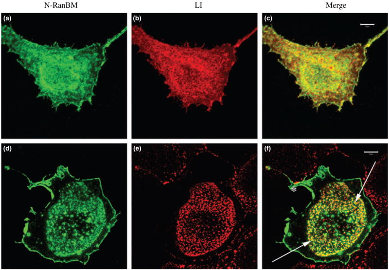
Fig. 3.

Colocalization of L1 and N-terminal region of RanBPM (N-RanBPM) after antibody-induced patching of L1. These confocal images represent Z-stacks through the cells, (a–c) COS-7 cells cotransfected with HA-tagged N-RanBPM (a) and L1 (b). Cells were fixed and stained without patching, (c) N-RanBPM (green) showed extensive colocalization with L1 (red). Bar, 10 μrn. (d–f) COS-7 cells cotransfected with HA-tagged N-RanBPM (d) and L1 (e). In this field only one cell expressed N-RanBPM while adjacent cells expressed some L1. Cells were patched with Ranti-L1 for 10 min before fixation. L1 (red) exhibited a patchy distribution in the center of the cell and N-RanBPM (green) was partially colocalized with L1 patches indicated with arrows. Some RanBPM remained at the perimeter of the cell (asterisk) and is not associated with L1. Bar, 10 μrn.
