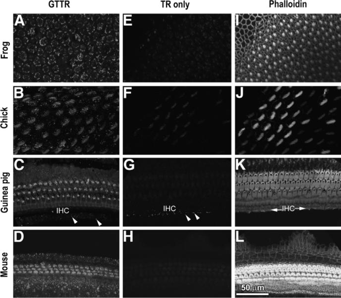
Fig. 1.

GTTR fluorescence in: (A) bullfrog saccule, (B) chick basilar papilla, (C) guinea pig cochlea and (D) 6-day-old murine cochlea 24 h after GT/GTTR injection (chick, 9 h). Panels (E–H) show negligible TR fluorescence in bullfrog saccule, chick basilar papilla, guinea pig and mouse cochleae, respectively, at equivalent time points and identical laser settings. (F) Weak, non-specific fluorescence is visible in chick stereociliary bundles, due to crosstalk from FITC-phalloidin-labeled stereocilia. (G) Weak autofluorescence (or aldehyde-induced fluorescence; arrowheads) is present in the inner sulcus region of the guinea pig, adjacent to the inner hair cell region (IHC in C, K). (I–L) Phalloidin-labeled sensory epithelia imaged in panels E–H, showing actiniferous regions at the lumenal surface. Scale bar applies to all images.
