
Fig. 4.
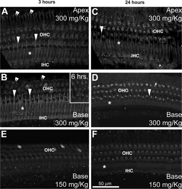
Guinea pig organ of Corti examined at 3 (A, B, E), and 24 h (C, D, F) after 300 mg/kg (A–D) or 150 mg/kg GT/GTTR injection (E, F). Three hours after injection, hair cells at the base of the cochlea (B) display slightly greater intensities of GTTR fluorescence compared to apical hair cells (A). This apparent gradient had disappeared 24 h post-injection (C, D). OHCs have greater fluorescence than IHCs. Note that at 3 h (A, B, E), only diffuse GTTR fluorescence can be observed in the organ of Corti (except for the punctate autofluorescence adjacent to IHCs). Sparse, punctate labeling is first seen in basal cochlear OHCs 6 h post-injection (B, inset), and all OHCs after 24 h (D, F). Note that the outer pillar cell phalanges (arrowheads) between the first row of OHCs, in the Deiters' cell phalanges of the third row (double arrowheads) and the inner pillar cell phalanges (*) are also diffusely labeled. Scale bar applies to all images.