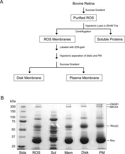
Fig. 2.
A, flow diagram showing the main steps in the purification and fractionation of bovine ROSs. B, Coomassie Blue-stained SDS-polyacrylamide gels of the various ROS fractions used in our proteomics studies. From left to right are molecular weight standards (Stds), ROS, soluble fraction (Sol), rod outer segment membranes (Mem), disk fraction (Disk), and enriched plasma membrane fraction (PM). Each lane was loaded with 30 μg of proteins. Rho, rhodopsin monomer; Rho(2), rhodopsin dimer, CNGB1, cyclic nucleotide-gated channel (B1 or β subunit).

