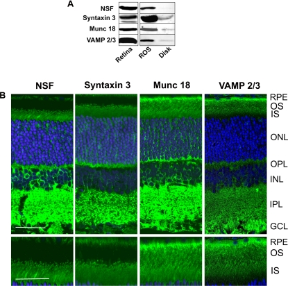
Fig. 6.
Detection and localization of SNARE proteins in the retina and ROSs. A, Western blots of retina, ROSs, and disks labeled with antibodies to NSF, syntaxin 3, Munc 18-1, and VAMP2/3. B, top panel, confocal scanning microscopy of rat retinal cryosections labeled with primary antibodies to NSF, syntaxin 3, Munc 18-1, and VAMP2/3 followed by a secondary antibody tagged with the Cy3 fluorescent dye (green) and counterstained with the DAPI nuclear dye (blue). Bar, 20 μm. Bottom panel, same as top panel except showing a restricted region of the outer and inner segments of photoreceptor cells at higher magnification. Bar, 10 μm. Similar labeling results were obtained with mouse retina. Retinal layers are: OS, outer segments; IS, inner segments; ONL, outer nuclear layer; OPL, outer plexiform layer; INL, inner nuclear layer; IPL, inner plexiform layer; and GCL, ganglion cell layer.

