
Figure 2. Diminished TGFβ Signaling in Human Genital SQCCs.
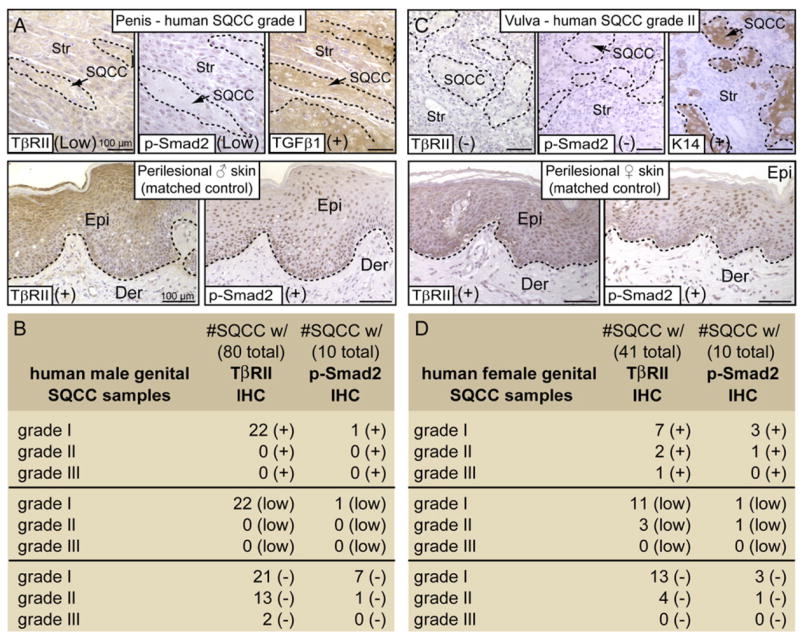
Male (B) and female (D) human genital SQCCs were scored according to their grade of severity (I-lowest to III-highest) and analyzed for TβRII, phosphorylated (activated) Smad2, TGFβ1 and/or K14 by immunohistochemistry (IHC). Examples of IHC staining from a male genital SQCC grade I (A) and from a female SQCC grade II (C) show reduced or no anti-TβRII, in contrast to perilesional, matched control skins. TβRII loss correlated with a lack of p-Smad2 staining in 10 out of 10 SQCC samples. TGFβ1 and K14 served as internal quality controls and were detected in TβRII-deficient human samples. Scoring: (+), positive staining; (−), negative staining; (low), reduced expression. Epi, epidermis; Der, dermis; Str, stroma. Lines encircle SQCCs in the top panels.