
Figure 6. Wound Closure Is Accelerated in cKO versus WT Animals.
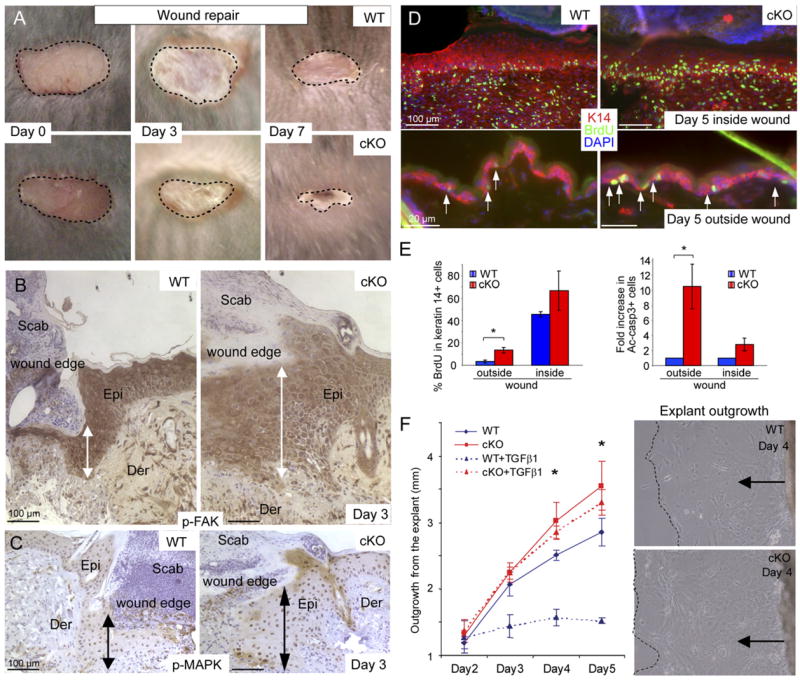
(A) Shown are representative examples at 0, 3, and 7 days after wounding.
(B and C) Immunohistochemistry reveals activated FAK and MAPK at the epidermal wound edge by day 3 after wounding. Note hyperthickening of wounded cKO skin. Epi, epidermis; Der, dermis.
(D and E) Quantification of proliferation and apoptosis inside and outside the wound area. Bar graphs depict mean ± SD. *p < 0.05.
(F) WT and cKO skin explant outgrowth in serum-containing media indicate accelerated epidermal outgrowth from cKO skin explants, a difference that becomes even more pronounced upon addition of active TGFβ (5 ng/ml) to the culture medium on day 2. Graph indicates the mean value of three independent experiments (±SD). *p < 0.03. Phase contrast images show extent of outgrowth at 4 days in the absence of active TGFβ .