
Figure 7. Elevated Cell Motility in the Absence of TbRII.
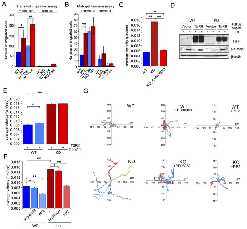
(A and B) Elevated cell motility in the absence of TβRII. Transwell migration assays were carried out on Boyden chambers ± coating with Matrigel ECM on the top chamber and in the absence and presence of fibroblast-conditioned medium, used as stimulus in the bottom chamber. Bar graphs depict means of 3 independent experiments ± SD. *p < 0.03. **p < 0.004. Note that KO MKs showed elevated migration and invasion over WT MKs, and Ha-Ras transformation enhanced these effects.
(C and D) Rescue experiment to show that the enhanced migratory ability of KO MKs is specifically attributable to TβRII loss. The CMV promoter was used to drive TβRII cDNA, which restored cell velocities to WT levels (C). Immunoblot shows that re-expression of TβRII in KO MKs restores TGFβ2 responsiveness (D).
(E) TGFβ1 treatment has only a slight effect on the velocity of WT MKs, which are still significantly less motile than KO MKs.
(F and G) Enhanced MAPK (Erk) and Src/FAK activities promote cell motility in KO MKs. (F) Cell velocities were measured ± 50 μM of the MEK1 inhibitor PD98059 and ± 5 μM of the Src inhibitor PP2, which also inhibits FAK activity. (C, E, and F) Thirty cells were tracked by phase-contrast microscopy with an acquisition rate of 1 frame/min over 12 hr. Bar graphs indicate average velocity ± SEM. *p < 0.05. **p < 0.0001. (G) Scatter plots depict the migration tracks of 5 representative cells for each condition.