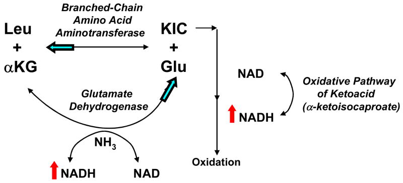
Figure 3. Schematic overview of glutamate metabolism.
Respiratory chain dysfunction presumably results in an increased NADH:NAD+ ratio (indicated by red arrows), which limits oxidation of ketoacids such as α-ketoisocaproate (KIC), the ketoacid of leucine. The result is greater accessibility of the ketoacid for conversion to its parent amino acid – in this illustration, leucine (Leu). A consequence would be accumulation of the parent amino acid and relative depletion of glutamate (Glu) (see Table 3). Some glutamate might be derived from reductive amination of −–ketoglutarate (αKG) in the glutamate dehydrogenase reaction, which is enhanced by an increased NADH:NAD+ ratio. However, this reaction is not sufficiently robust to prevent an overall diminution of the glutamate concentration and a marked increase in the ratio of leucine (and other amino acids) to glutamate (Table 3). This suggests that levels of amino acids such as leucine, which are formed from transamination reactions with glutamate, may be sensitive indicators of an altered redox state. Postulated direction of equilibrium reaction alterations occurring in primary respiratory chain dysfunction are indicated by blue arrows.

