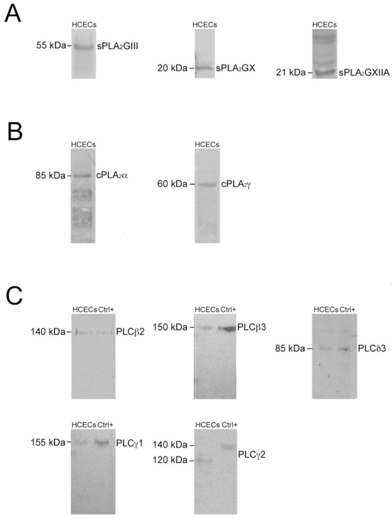
Figure 3.

Western blot analysis of phospholipases in crude extracts from HCECs. Western blot analyses were performed using the (A) sPLA2, (B) cPLA2, and (C) PLC antibodies shown (Table 2). Left: molecular mass of the expected proteins. Ctrl+, positive controls RAW 264.7 for PLCs β2 and δ3, A-431 for PLCs β3 and γ1, and MCF7 for PLCγ2.
