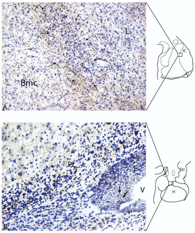
Figure 3.
(A) Photomicrograph of 5-HTT-positive fibers (brown) densely innervating an intercalated island (arrows). The basal nucleus to the left, and lateral nucleus to the right, both contain a lighter innervation by 5-HTT labeled fibers. The island is interposed between the basal and lateral nucleus which have larger, less densely packed cells (boxed area of the schematic shows position of the intercalated island). (B) Photomicrograph of 5-HTT labeled fibers (brown) in a Nissl stained caudal section through the amygdala. Labeled fibers in the paralaminar nucleus near the lateral ventricle extend to the ependymal lining (arrows). Bmc, basal nucleus, magnocellular subdivision; PL, paralaminar nucleus; V, ventricle; E, ependymal lining of ventricle; L, lateral nucleus; H, hippocampus; IC, intercalated cell islands; Str, striatum.

