
FIG. 1.
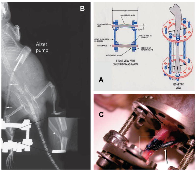
(A) Fixator used for DO in the mouse model and how it is applied to the tibia around the osteotomy site. (B) As shown in this whole animal radiograph, Alzet Model 1002 miniosmotic pumps were inserted subcutaneously, and infusion tubing (arrows) was secured and routed subcutaneously from the pump to the distraction site for local delivery. (Inset) The catheter’s opening was placed adjacent to the osteotomy. (C) Methylene blue was infused for 14 days by local infusion. At death, surrounding soft tissues were dissected away from the surgical site, exposing the distraction gap, adjacent bone, and the infusion catheter tip. Blue dye was seen only in the distraction gap (boxed area) and in the catheter tip (arrow).