
FIG. 3.
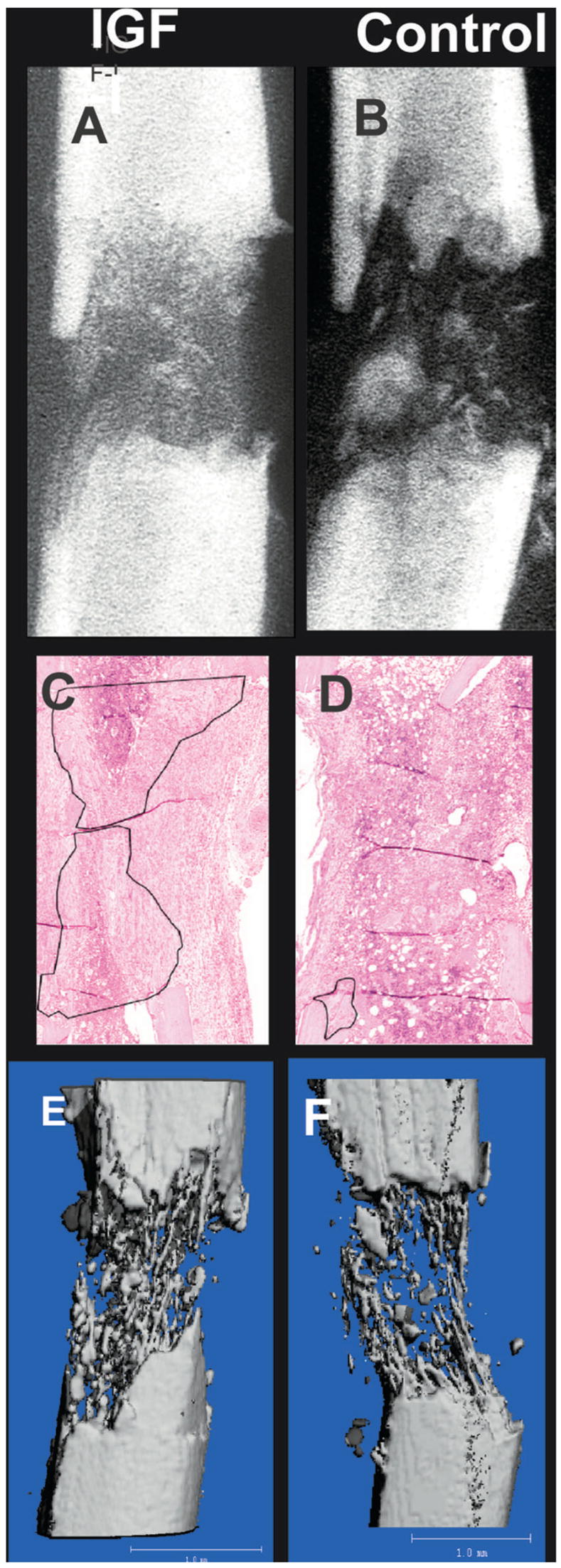
Representative radiographs of a distraction gap from (A) an rhIGF-I–treated mouse and (B) a vehicle-treated mouse (control). Histological sections from representative animals treated (C) with or (D) without rhIGF-I are presented, showing new bone formation outlined in black. (E and F) Effects of rhIGF-I infusion on bone formation was refined further using μCT. Representative μCT reconstructions of distraction gaps from (E) an rhIGF-I–treated and (F) a vehicle-treated mouse are shown.