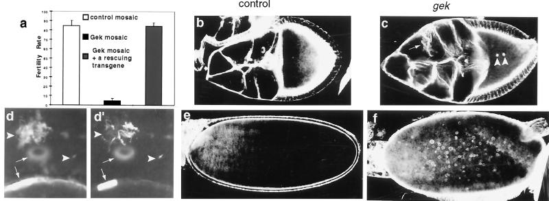
Figure 5.
gek mutants are defective in oogenesis and in actin cytoskeletal organization in egg chambers. (a) Fertility rate of eggs laid from control mosaic females (open bar, genotype: yw,P[hsFLP]/+;FRT,P[ovoD1]/FRT), gek mosaic females (black bar, genotype: yw,P[hsFLP]/+;FRT, P[ovoD1]/FRT,gekD23) and gek mosaic females in the presence of one copy of a rescuing transgene (gray bar, genotype: yw,P[hsFLP]/+;FRT,P[ovoD1]/FRT,gekD23;T35/+). (b and c) Phalloidin staining of stage 9 control (b) and gek (c) egg chambers. (d and d′) High-magnification images in nurse cells of a stage 9 gek mosaic egg chamber double-labeled with phalloidin (d) and anti-Hts (d′). In gek mutant clones, the cortical actin surrounding the nurse cells is disorganized (arrow in c) compared with the smooth structure in control mosaics (b). Ectopic actin polymerization occurs in oocytes (arrowheads in c) and in nurse cells (arrowheads in d). Such ectopic F-actin structures in nurse cells also are recognized by antibody to Hts (arrowheads in d′). Small arrows indicate the ring canals. (e and f) Phalloidin staining of stage 12 control (e) and gek (f) oocytes. In late-stage oocytes ectopic F-actin forms numerous spheres surrounding the yolk granules in gek mosaics (f). No such actin spheres are found in oocytes of control mosaics (e). Genotypes of control and gek mosaics in b–f are the same as in a.

