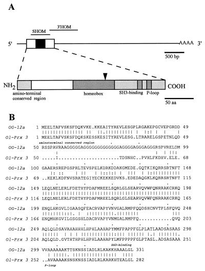
Figure 1.
(A) Schematic representations of Ol-Prx 3 cDNA (Upper, clone E) and putative protein structures (Lower). The open reading frame (box), homeobox (black box), and poly(A) tail locations are given in the cDNA drawing. Probes used for Northern blotting and in situ hybridizations are shown above the cDNA structure. Shaded boxes in the protein drawing show regions conserved with OG-12. Names of the conserved amino-terminal, homeodomain, SH3-binding, and P-loop domains are given below their respective locations. The oligonucleotide involved in library screening is indicated by an arrow. (B) Comparison of the amino acid sequence encoded by medaka Ol-Prx 3 and that of mouse OG-12. The amino-terminal, SH3-binding, and P-loop domains are named below. Double dots indicate conservative amino acid changes in OG-12 vs. Ol-PRX 3 proteins.

