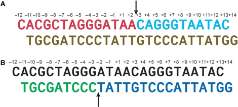
Figure 1.
Nicked DNA duplexes used in crystallization studies. (A) A DNA duplex containing a nicked top strand assembled from two oligonucleotides (colored red and cyan) and an intact bottom strand oligonucleotide (brown). (B) A DNA duplex containing a nicked bottom strand assembled from two oligonucleotides (colored green and dark blue) and an intact top strand (colored black). The arrows indicate the cleavage sites on the top and bottom strands.

